

BD
Biotin Anti-Human and Viral IL-10 JES3-12G8
SKU · 554499
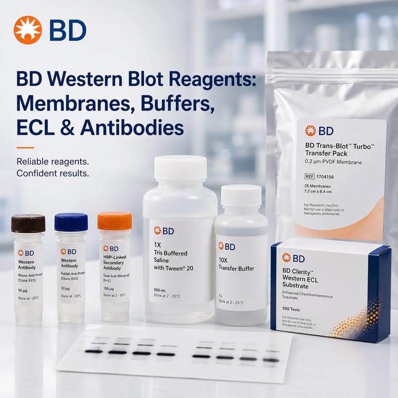
From membrane selection to signal detection, BD Western Blotting & Molecular Reagents give you a cohesive, validated toolkit for reliable blots. This Collection page helps you choose faster with scenario-based guides and side-by-side comparisons. As an authorized distributor, Iright supplies a wide range of BD membranes, buffers, antibodies, and detection substrates.
Choosing the right membrane, buffer system, secondary antibody, and substrate determines whether your bands are clear, linear, and publication-ready. This section explains where BD’s portfolio fits in a full Western blot workflow so you can match products to your samples and imaging systems with confidence.
What this series enables
Each product family maps to a specific performance need—binding strength, background suppression, or sensitivity. Use the summaries below to shortlist families before you dive into the selection and comparison tables.
Membranes (PVDF / NC)
PVDF offers high protein-binding capacity and solvent compatibility; it’s ideal for low-abundance targets or when reprobing is planned. NC membranes provide fast binding and low autofluorescence, favored for routine targets and clean backgrounds. Select pore size by target molecular weight.
Buffers (Running / Transfer / Blocking / Wash / Stripping)
Running and transfer buffers maintain protein integrity and mobility; methanol in transfer mixes improves binding on PVDF. Blocking buffers (protein- or polymer-based) minimize nonspecific binding; choose by membrane type and antibody host. Wash buffers balance stringency and epitope retention. Stripping buffers enable antibody removal without compromising target proteins.
Secondary Antibodies (HRP / AP / Fluorophore-labeled)
Species-specific secondaries matched to your primary host are available in HRP or AP conjugates for chemiluminescence, and in multiple fluorophores for multiplex detection. Affinity-purified options enhance specificity and reduce background.

ECL Chemiluminescent Substrates
From standard sensitivity to ultra-sensitive, ECL formulations differ in signal duration, intensity, and dynamic range. Pair the substrate to the expected target abundance and your camera’s detection limits.
Selection starts with your biological context—sample type, target abundance, and detection method. Use the table as a fast path to an initial kit of membrane, buffers, secondaries, and substrate; then confirm fine-grain parameters in the comparison section.
| Scenario | Membrane (Type) | Transfer Buffer | Blocking Buffer | Secondary (Host/Label) | Substrate/Channel | Notes |
|---|---|---|---|---|---|---|
| Low-abundance nuclear protein | PVDF (0.2 μm) | Methanol-containing (cold) | Protein-free polymer | Anti-rabbit HRP | High-sensitivity ECL | Extend blocking; minimize over-exposure |
| Routine cytosolic target | NC (0.45 μm) | Standard Tris-glycine | BSA-based | Anti-mouse HRP | Standard ECL | Shorter exposure for linearity |
| Multiplex phospho/total pair | PVDF (0.2 μm) | Methanol-containing | Synthetic polymer | Anti-rabbit / anti-mouse fluorophores | 700/800 nm channels | Control cross-talk; sequential incubation |
| High-lipid tissue lysate | PVDF (0.45 μm) | Methanol-containing, chilled | Casein-based | Anti-rabbit HRP | Enhanced ECL | Prolonged washes; add detergent to TBST |
| Reprobe same target set | PVDF (0.2 μm) | Standard | Polymer blocker | HRP | Standard ECL | Gentle stripping; validate housekeeping band |
Use this table to compare BD’s most frequently used Western blot detection reagents and WB-validated antibodies. Pack sizes/usage notes follow BD datasheets; always confirm the latest specs on the product page before ordering.
| SKU | Product Name | Type | Detection | Pack Size |
|---|---|---|---|---|
| 554002 | BD Pharmingen™ HRP Goat Anti-Mouse Ig | Secondary antibody | HRP → ECL | 1 mL |
| 554021 | BD Pharmingen™ HRP Goat Anti-Rabbit Ig | Secondary antibody | HRP → ECL | 1 mL |
| 554017 | BD Pharmingen™ HRP Goat Anti-Rat Ig | Secondary antibody | HRP → ECL | 1 mL |
| 550946 | BD Pharmingen™ Streptavidin-HRP | Conjugate | HRP → ECL | 1 mL |
| 554066 | BD Pharmingen™ Streptavidin-HRP | Conjugate | HRP → ECL | 1 mL |
| 550337 | Biotin Goat Anti-Mouse Ig (Multiple Adsorption) | Linker (biotinylated) | Biotin ↔ SA-HRP/Fluor | 0.25 mg |
| 612657 | Purified Mouse Anti-Actin Ab-5 (Clone C4) | Primary antibody (WB control) | HRP/Fluor (via secondary) | 150 µg |
| 610340 | BD Transduction Laboratories™ Purified Mouse Anti-Ran | Primary antibody | HRP/Fluor (via secondary) | 50 µg |
| 610088 | BD Transduction Laboratories™ Purified Mouse Anti-FAK | Primary antibody | HRP/Fluor (via secondary) | 150 µg |
| 565781 | BD Pharmingen™ Purified Mouse Anti-Human Perlecan | Primary antibody | HRP/Fluor (via secondary) | 0.1 mg |
Even with the right components, small choices—like blocker type or wash stringency—change outcomes. Use the following principles to lock in a low background and a usable linear range across different imaging setups.
Membrane × Blocker
Detection Chemistry
Washes & Stringency
A concise, repeatable protocol reduces variability across operators. Adapt times to your gel percentage, target size, and imaging hardware; keep notes on exposure to preserve linearity for densitometry.
Mini Protocol (outline)
Troubleshooting quick cues
High background: Switch to polymer blocker; lengthen washes; reduce antibody concentration.
Weak signal: Use higher-sensitivity ECL; ensure transfer efficiency; confirm primary specificity.
Non-specific bands: Increase stringency; shorten primary incubation; validate species cross-reactivity.
Uneven bands: Level gel/transfer stack; reduce current; ensure uniform membrane contact.
Your team will likely search for practical answers during setup or when a blot misbehaves. These concise responses keep the page useful and capture long-tail queries without keyword stuffing.
Q1: ECL or fluorescence for quantitative blots?
If you need multiplexing or very wide linearity, fluorescence excels—especially with two-color phospho/total workflows. For routine targets, HRP + ECL is fast and sensitive; choose a high-sensitivity ECL only when truly necessary to avoid saturation.
Q2: PVDF vs NC—how should I choose?
Use PVDF for low-abundance or hydrophobic/membrane proteins and for repeated stripping/reprobing. Choose NC for clean backgrounds with mid-abundance proteins and when you plan fluorescent detection with minimal autofluorescence.
Q3: What blocking buffer reduces background most reliably?
Match blocker to membrane and epitope: polymer or casein blockers tame hydrophobic binding on PVDF; BSA is often sufficient on NC. Avoid milk when probing phospho-proteins to prevent casein cross-reactivity.
Q4: How do I keep the signal within a linear range?
Lower ECL strength or reduce exposure; titrate primary and secondary antibodies; ensure transfer is complete but not excessive. Capture several exposures and pick the one that keeps housekeeping and target bands unsaturated.
Q5: Can I strip and reprobe the same membrane?
Yes—use mild stripping buffers and re-block thoroughly. Validate that housekeeping bands remain intact; if targets are labile, consider running duplicate gels instead of aggressive stripping.
Filter this collection by brand or tags.