Description
The K10-895.12.50 monoclonal antibody recognizes the phosphorylated serine 529 (pS529) in the transactivation domain of the human NF-κB p65 subunit. Nuclear factor κB (NF-κB) is a ubiquitously expressed transcription factor that regulates the expression of many other genes. It is crucial for cellular responses to a variety of stimuli including stress and microbial pathogens that lead to immunity, inflammation, proliferation, differentiation, survival, apoptosis, and tumorigenesis. The most studied NF-κB complex consists of the p50 (also known as NF-κB1) and p65 (also known as REL-A) subunits, both containing a 300-amino acid region with homology to the Rel proto-oncogene product (RH domain). The RH domain contains motifs for dimerization, nuclear localization, and binding to specific DNA sequences. In addition to the RH domain, the p65 subunit contains the transactivation domain, which is responsible for the interaction with the inhibitor IκB and which contains phosphorylation sites. In most cell types, the p50/p65 heterodimer is located within the cytoplasm complexed to IκB. This complex prevents nuclear translocation and activity of NF-κB. In response to stimuli such as cytokines, LPS, DNA damage, and microbial infections, IκB is phosphorylated at critical residues. This phosphorylation induces dissociation of the IκB/NF-κB complex, allowing the free heterodimeric NF-κB to translocate to the nucleus. Furthermore, optimal activation of NF-κB requires phosphorylation in the transactivation domain of p65. In the nucleus, activated NF-κB dimers bind to the κB sites within promoters and enhancers and function as transcriptional regulators.
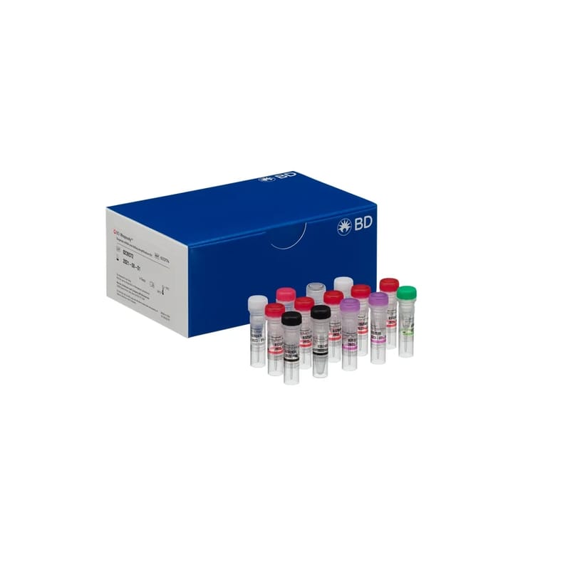
This antibody conjugate is suitable for intracellular staining of human cell lines and peripheral blood mononuclear cells using BD Cytofix™ Fixation Buffer. Any of the three BD Phosflow™ permeabilization buffers may be used. For more information and suggested protocols, investigators are encouraged to visit http://www.bdbiosciences.com/research/ics/resources/index.jsp
Store undiluted at 4°C and protected from prolonged exposure to light. Do not freeze. The monoclonal antibody was purified from tissue culture supernatant or ascites by affinity chromatography. The antibody was conjugated to Alexa Fluor® 647 under optimum conditions, and unreacted Alexa Fluor® 647 was removed.